-
STAR-D officially selected for the REXUS/BEXUS programme
I am delighted to be mentoring a team (Star-D) taking part in the REXUS program to develop an experiment for a sounding rocket, including our own Dom Alderson. They have now officially been selected! I copy their statement below:
“We are delighted to announce that STAR-D has been officially selected to take part in the REXUS/BEXUS programme.
This selection marks a historic first: it is the very first time ever that the Pôle Léonard de Vinci, together with AgroParisTech, Newcastle University, and the University of Vienna, will participate in a mission of this scale with European Space Agency – ESA.
STAR-D is also the only French team selected for this REXUS/BEXUS cycle.
We are extremely grateful to the selection committee for recognizing our work, and proud to be supported by our mentors Didier Gossard and Adam Wollman, as well as by LéoFly, the aerospace association carrying the project.
We are now entering the core phase of the programme.
Next step: the student training week and the Preliminary Design Review at the Esrange Space Center in Sweden.
Stay tuned for the next chapter 🚀
STAR-D is a LéoFly project studying phagocytosis in microgravity, using a modified OpenFlexure microscope combined with a custom microfluidic system, flown aboard a suborbital rocket.
About REXUS/BEXUS
REXUS/BEXUS is a student programme enabling experiments to fly on sounding rockets and stratospheric balloons. It was co-created by the Rymdstyrelsen and German Aerospace Center (DLR), and is now run in collaboration with ESA, MORABA, SSC – Swedish Space Corporation, and ZARM.”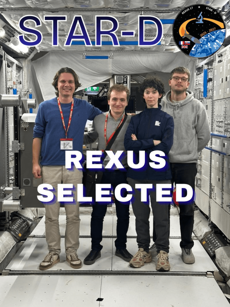
-
FlightScope: An open source microscopy platform for microgravity research
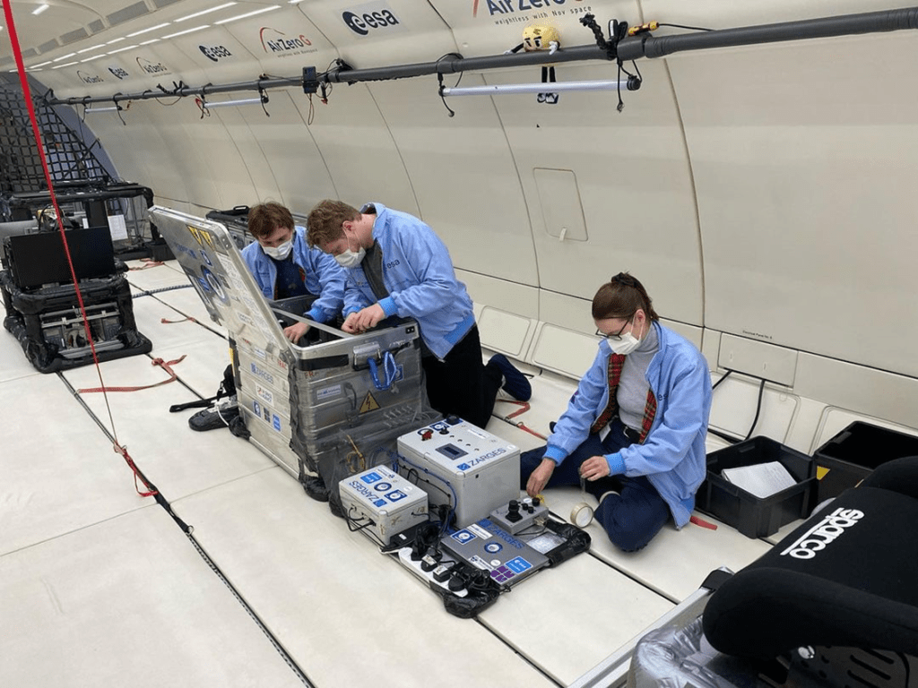
With planned missions to the Moon and Mars, understanding the impact of microgravity on biological organisms has never been more important. Parabolic flights offer short periods of simulated microgravity using modified commercial airliners. However, studying live cells on parabolic flights is a major challenge due to intense aircraft vibration and very short 15-20 second windows of weightlessness. To solve this, we developed FlightScope, a new microscopy and microfluidics platform designed to study dynamic cellular processes in real-time during these flights, recently published in npj Microgravity (pre-print also available).
We engineered FlightScope to be a vibration-resistant fluorescence microscope that could withstand the tough conditions on the plane. Its most unique feature is a bespoke microfluidics system that allows us to inject substances into our cell samples; observe their behaviour live, during the rapid changes in gravity; and quickly change sample between parabolas. By using an open-source design (SQUID microscope) and 3D-printed parts, we also made our platform cost-effective, costing under $10,000 to build.
We put our system to the ultimate test on board a European Space Agency (ESA) parabolic flight. During the flight, we successfully obtained high-quality images of live yeast cells. Using our integrated microfluidics, we injected a fluorescent glucose solution and recorded the cells taking it up in real-time as we transitioned from microgravity to hypergravity. The successful performance of FlightScope provides an important proof-of-principle that opens the way for future investigations into cell biology in space. Furthermore, its rugged and contained design makes FlightScope a versatile tool for microscopy in other extreme environments here on Earth. We are actively working with groups on new microgravity experiments but always looking for new collaborators
-
Fully Funded PhDs available in the Wollman lab on the new NEEDL DTP
Two related PhD projects to advertise in my lab and Josana Rodriguez lab at the Newcastle Biosciences Institute. 4 years fully funded.
Understanding cellular rapid shape shift using molecular rulers
https://lnkd.in/efwDFmKz
The role of membrane dynamics in the polarisation of cells during embryo development
https://lnkd.in/eZPQNHmj
Both using cutting-edge microscopy, AI and genetics. Full training given. All scientific backgrounds considered, please get in touch if interested. -
PhD Studentship Available on the BBSRC NLD DTP
Another fully funded PhD project is available in the lab through the BBSRC NLD doctoral training partnership. Come join the lab using CRISPR/Cas9 to fluorescently label specific sections of the genome and observe gene regulation in real time with single-molecule lightsheet microscopy. Biology, physics or any background in between please apply!
-
Paper accepted in JMB
Tetrameric UvrD helicase is located at the E. coli replisome due to frequent replication blocks

The sister paper to our study on the Rep helicase. Using dual colour live cell single-molecule fluorescence microscopy, we investigated how E.coli’s other accessory helicase, UvrD, functions. We found that differently to Rep, UvrD, is not recruited to the replisome by a specific replisome partner. Rather the replisome encounters so many other blocks to replication which recruit UvrD that there is almost always UvrD present at the replication fork.
-
Two MRC funded PhD studentships available in the lab through the DiMeN DTP!
Work with us to develop new treatments for Alzheimer’s disease by reducing inflammation.
Investigating the adaptive resistance of Pseudomonas aeruginosa raised from antibiotic persistence
Use super-resolution and atomic force microscopy to understand how bacteria develop antibiotic resistance
-
New paper – Reduced binding of apoE4 to complement factor H promotes amyloid-β oligomerization and neuroinflammation
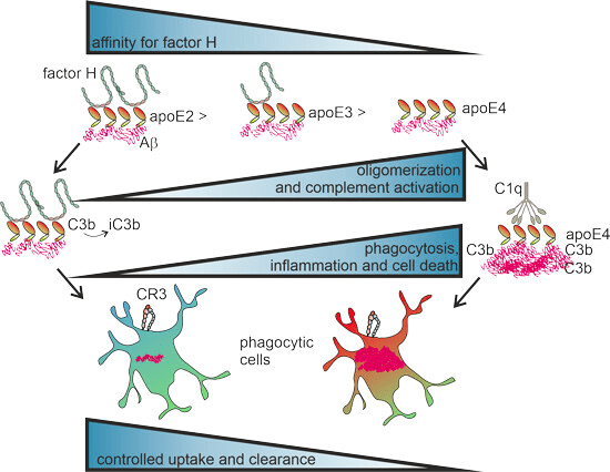
New paper with our collaborator, Dr Karita Haapasalo at the University of Helsinki. In the paper, we elucidate the molecular mechanism of a genetic risk factor for Alzheimer’s disease. We show how the complement system interacts with apolipoprotein E (ApoE) to clear amyloid beta plaques and how this is impaired in the ApoE4 variant associated with late-onset Alzheimer’s. At Newcastle, we used single-molecule microscopy to visualise molecular ApoE-amyloid complexes directly and developed bespoke image analysis software to characterise these complexes in patient brain biopsy samples. (https://www.embopress.org/doi/full/10.15252/embr.202256467)
-
Team SUGAR with the European Space Agency
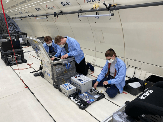
The lab was lucky enough to get to work with the European Space Agency (ESA) developing a microscope – Gravityscope – to image cell signalling in live cells on board a parabolic flight. Our team – SUGAR (Saccharomyces Cerevisiae Uptake of Glucose and Real-time imaging) spent 12 months developing Gravityscope, a robust fluorescence microscope incorporating microfluidics, to image uptake of fluorescently tagged glucose in live yeast cells as plane gravity switched from normal to microgravity to hypergravity and back again – all over 90s – no mean feat!
-
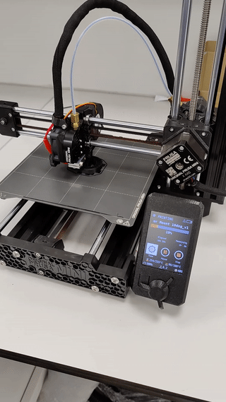
New lab 3D printer!
-
Wollman Lab
We have launched our new lab website!
-
Subscribe
Subscribed
Already have a WordPress.com account? Log in now.
